The ophthalmologist will check to see if the eye moves as it should and if there are any vision problems.
Ct scan obital floor plate.
A 3 d ct of unilateral clinical anophthalmia with hypoplasia of the ipsilateral bone.
They will take pictures of the eye and the eye socket including x rays and ct scans.
The use of a polydioxanone pds plate for orbital reconstruction was evaluated in 20 patients with various traumatic defects of the orbital floor.
Coronal ct shows ablation of the greater wing of the sphenoid bone od with herniation of the temporal lobe into the orbit causing hypoglobus and pulsatile proptosis.
Radiographic analysis showed that in 12 of the 13 patients there was new bone in the orbital floor.
We use ct scan data to design the titantium implants to approximate the anatomy of the orbital floor and medial wall.
These plates consist of implants that closely approximate the topographical anatomy of the human orbital floor and medial wall and are intended for use in a selective craniomaxillofacial trauma.
This is typically caused by a direct blow to the central orbit from a fist or ball.
To check for an orbital fracture an ophthalmologist will examine the eye and the area around it.
Plate designed from ct scan data to approximate solutions anatomy of orbital floor and medial wall orbital floor.
This x ray shows the classic transition zone.
Plate borders medial wall orbital floor designed from ct scan data the three dimensional implants closely approximate the topographical anatomy of the hu man orbital floor and medial wall to provide accurate recon struction even after significant two wall fractures 5 6 preformed three dimensional shape.
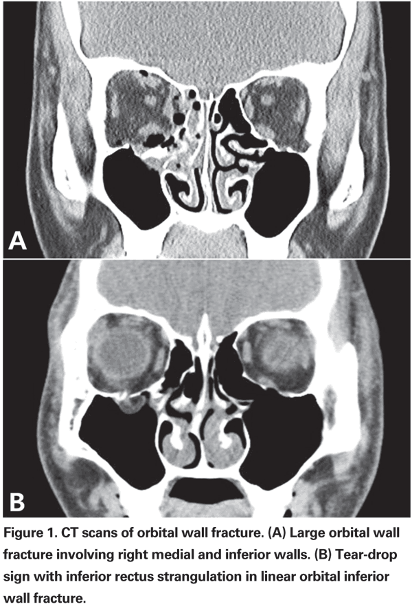
Orbital blowout fractures occur when there is a fracture of one of the walls of orbit but the orbital rim remains intact.
The follow up time was 9 to 45 months mean 20 4 months.
The floor is likely to collapse because the bones of the roof and lateral walls are robust.
Coronal slice of a postoperative ct scan taken after transconjunctival repair of the complete left medial orbital wall and orbital floor.
The matrixmidface preformed orbital plates are designed from ct scan data.
An orbital blowout fracture is a traumatic deformity of the orbital floor or medial wall typically resulting from impact of a blunt object larger than the orbital aperture or eye socket most commonly the inferior orbital wall i e.
The overlying colored line in the medial wall and orbital floor area indicate the preoperative virtual planning that is superimposed on the mesh reconstructed area.
We then cover the implant with our proven medpor biomaterial to minimize sharp edges even if the plate requires modification.
A ct scan was obtained in 13 patients.